In all living organisms, proteins are the machineries that carry out all the biological processes inside and outside the cells. The understanding of their structure is an important step in figuring out how they work and why an alteration in their structure, and in turn their function, results in disease. Indeed, the protein functions are strictly related to their structures even when proteins are without a defined three-dimensional structure, as in the case of intrinsically disorder proteins. But how do scientists unravel the structures of such macromolecules?
Structural biology is the answer. Structural biology is a branch of biochemistry that studies the structure of macromolecules, such as proteins and nucleic acids. Using and integrating different techniques, structural biology allows the determination of the atomic-detail structure of molecules that otherwise would not be visible. The most commonly used methods are X‐ray crystallography, nuclear magnetic resonance (NMR), and cryo‐electron microscopy (EM). Structural studies can impact on our understanding of neurodegenerative diseases. Amyotrophic lateral sclerosis, Parkinson's disease, and Alzheimer's disease among others, are characterised by the gradual loss of neuronal function due to protein aggregation and/or dysfunction. Clarifying the pathophysiological mechanisms behind these diseases is the first step towards the development of effective drugs that can prevent or treat such diseases.
X-Ray Crystallography: the old but gold method In 1953, Kendrew and Perutz determined the crystal structure of myoglobin and haemoglobin. Since then, X‐ray crystallography has been the dominant method in structural biology research, with the deposition of thousands of protein structures per year. Using X-ray beams and packing the biomolecules in crystals, X-ray crystallography gives a “photograph” at atomic resolution of the molecules in the crystal lattice. All the structural details are revealed: from structure interfaces to protein-protein interactions, as well as the binding sites of small molecules. For example, Wherman et al. determined the crystal structure of the extracellular domain of TrkA in complex with NGF, elucidating the residues involved in the interaction with the neurotrophin and providing a model for molecular docking in receptor-based drug design.
Crystal structure of the TrkA extracellular domain in complex with NGF. Image of 2IFG (Wehrman et al. (2007) Structural and mechanistic insights into nerve growth factor interac-tion with TrkA and p75 receptors”. Neuron 53:25-38) creat-ed with PyMol Molecular Graphics System, Version 2.0 Schrödinger, LLC.
Nuclear Magnetic Resonance Spectroscopy NMR spectroscopy provides protein three-dimensional structures at high resolution, as well as information on conformational dynamics and interactions that take place both in solution and in living cells. Thus, proteins that are too dynamic, disordered or not amenable to crystallisation can be examined by NMR spectroscopy. However, the application of this technique is limited to proteins with molecular weights of less than 50 kDa.
In addition, NMR spectroscopy is a powerful tool in screening the binding of compounds to target proteins for drug discovery research. Recently, Franco et al. determined the first NMR structure of the TrkA transmembrane domain (TM), showing the dimer length, the angle formed between the two helices, and the contact surface areas characterized by a conserved sequence motif. This information is fundamental to understanding the mechanism underlying receptor activation upon NGF binding, laying the groundwork for studying molecules that may trigger the receptor activation.
NMR structure of TrkA-TM dimers. Image of 2N90 (Franco et al. (2020) Structural basis of the transmembrane domain dimeriza-tion and rotation in the activation mechanism of the TRKA re-ceptor by nerve growth factor. Journal of Biolog-ical Chemistry 295:275-286)) created with PyMol Molecular Graphics System, Version 2.0 Schrödinger, LLC.
Single-Molecule Cryo-Electron Microscopy The structure of large macromolecular complexes or proteins that are difficult to crystallise can be determined by cryo-electron microscopy (cryo-EM). Cryo-EM uses a beam of electrons, whose wavelength is shorter than that of light, enabling the atomic image of molecules at high resolution to be determined. With the advent of new detectors and developments in sample preparation (protein vitrification), the use of this technique has exploded in the last few years. An example of the advantage of Cryo-EM for studying neurodegenerative diseases is the structure of γ-secretase, a multi-subunit complex of about 270 kDa involved in the formation of amyloid plaques in Alzheimer’s disease (AD). In AD brains, γ-secretase cleaves amyloid precursor protein (APP), generating peptides prone to aggregation and forming amyloid plaques.
In 2019, Zhou et al. have determined the cryo-EM structure of the γ-secretase in complex with the APP fragment, providing a model that is useful, not only to better understand the disease progress, but also for the rational design of substrate-specific inhibitors.
Cryo-EM density map of human γ-secretase (grey) in com-plex with APP fragment (marine). Image of 6IYC (R. Zhou et al. (2019) Recognition of the amyloid precursor protein by human γ-secretase. Science 363(6428)) created with PyMol Molecu-lar Graphics System, Version 2.0 Schrödinger, LLC.
Structural biology in the EuroNeurotrophin Network 
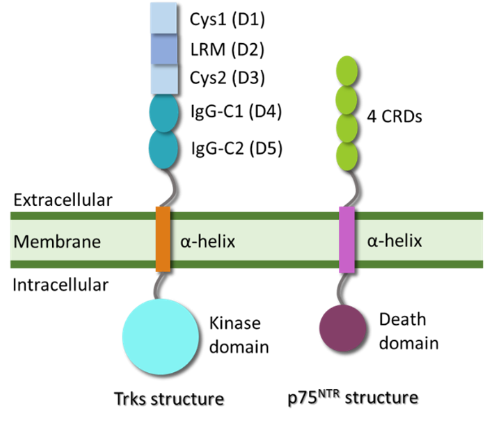
Within the “EuroNeurotrophin” network, we aim to crystallise and determine the structures of neurotrophin receptors in complex with small molecules by X-ray crystallography, in order to drive the optimization of lead compounds. Also, we want to integrate this structural information with functional studies by biophysical methods.
Federica is hosted at the University of Siena. Her research activities are focused on the production and crystallization of neurotrophin receptors in complex with selected compounds. The structural studies will clarify the determinants for ligands binding and hence drive receptor-based drug design.